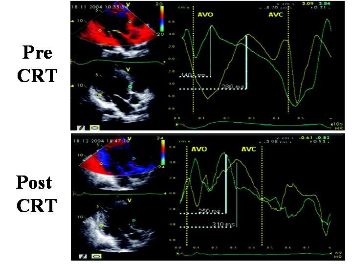
Figure 7

Sample of time to peak (Ts) measured by off-line color TVI pre and post CRT at the level of anterior septum and postero-lateral wall, in apical 5-chamber view. Aortic valve opening (AVO) and closure (AVC) are derived by previous placement of markers on the onset and the end of LV Doppler outflow. Pre-CRT Ts of postero-lateral wall (upper panel) is clearly delayed in comparison to Ts of anterior septum. Post-CRT the time duration of Ts between the two walls is clearly shortened (lower panel). (Modified from Innelli P et al, Echocardiography 2006;23:709–716)