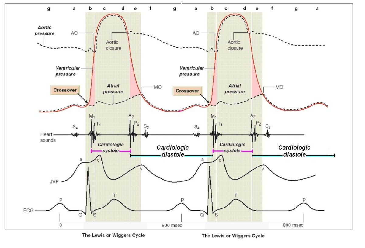
Figure 1

The mechanical events in the cardiac cycle, first assembled by Lewis in 1920 but first conceived by Wiggers in 1915. Cycle length of 800 milliseconds for 75 beats/min. Cardiological systole is demarcated by the interval between the first and the second heart sounds, lasting from the first heart sound to the closure of the aortic valve. The remainder of the cardiac cycle automatically becomes cardiological diastole. Left ventricular contraction: isovolumic contraction (b); maximal ejection (c). Let ventricular relaxation: start of relaxation and reduced ejection (d); isovolumic relaxation (e); LV filling rapid phase (f); slow LV filling (diastasis) (g); atrial systole or booster (a). Mitral valve closure occurs after the crossover point of atrial and ventricular pressures at the start of systole. A2 = aortic valve closure, aortic component of second sound; AO = aortic valve opening, normally inaudible; ECG = electrocardiogram; JVP = jugular venous pressure; M1 = mitral component of first sound at time of mitral valve closure; MO = mitral valve opening, may be audible in mitral stenosis as the opening snap; P2 = pulmonary component of second sound, pulmonary valve closure; S3 = third heart sound; S4 = fourth heart sound; T1 = tricuspid valve closure, second component of first heart sound. Modified from Opie LH. Mechanisms of cardiac contraction and relaxation. In: Braunwald E, Zipes DP, Libby P, Bonow RO, eds. Heart Disease. 7th ed. WB Saunders Company 2005, Chap.19:457–489, page 475.